Ear Reconstruction
When an ear malformation is visibly obvious, it can be a strain to live with. Many sufferers experience low self-esteem and the malformation makes them feel uneasy in their relationships with other people. This holds true for people of all ages. On the other hand, there are many sufferers, who are not affected in this way and consequently they don’t wish ear reconstruction surgery. These different personal perceptions are a central element of the consultation process.
Another important aspect in ear reconstruction is the age of the patient. Should reconstruction take place before a child is aware on their own malformation? Or, when the child is more mature and has already formed their own opinion about whether or not they want ear reconstruction? This will be discussed in detail during a consultation. Moreover, we will inform you about the advantages and disadvantages of the different treatment options. Therefore we would like to put each and every patient in the position to be able to take the decision on their own, armed with full information.
Ear reconstruction using 3D SuPor®
An extraordinary and unique procedure is the ear reconstruction made of 3D SuPor®. In this process, we use an individual, realistic 3-dimensional biocompatible synthetic framework made of polyethylene that reflects the patient’s own anatomy. It has become known as the “3D Lewin Ear” or “PIER”. This scaffold is encased in what is called a “fascial flap”. Fascia is a tissue layer of the scalp that is supplied with blood by its own vessels. The wrapped polyethylene scaffold is covered with skin grafts from the second ear or upper arm.


One advantage of this method is that only one operation is required and there is no need to remove rib cartilage. The scanning of the healthy ear with a laser scanner and the individual manufacturing process of the 3D SuPor® ear allows the reproduction of the complex and fine anatomy of the human ear to an unprecedented extent. This operation can be performed from the age of four to six years.
Ear Reconstruction using Rib Cartilage
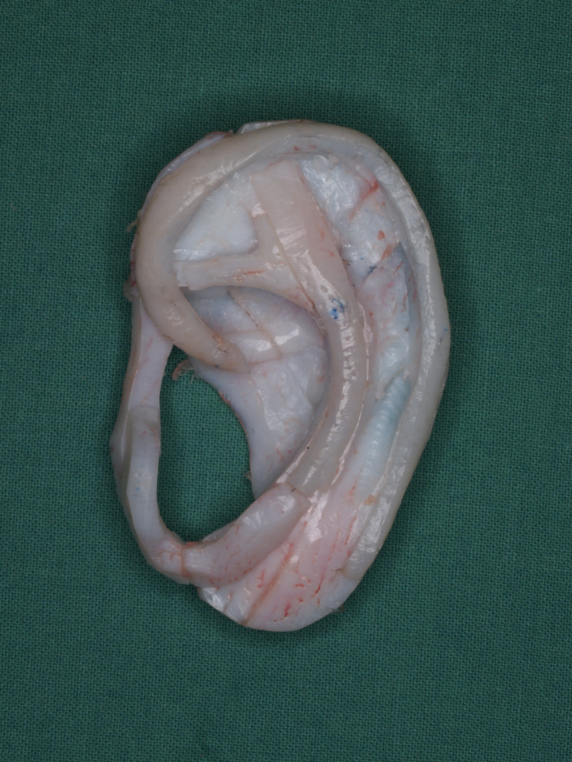
For ear reconstruction using rib cartilage, material from the patient’s own body is exclusively used. This method has been successfully used for decades and includes two operations with a gap of three months between them. When performed on children these operations are performed from the age of about eight to ten years.
During the first operation rib cartilage is harvested. Long-term pain or deformities of the thorax as a result of the operation are rare. A three-dimensional ear scaffold is created from the harvested cartilage. Where the patient has a normal second ear, this is used as a template. A pocket of skin is prepared for the ear scaffold and the lower part of the existing malformed external ear is used to create a new ear lobe. In this procedure, the lower part of the existing malformed pinna is used as a new earlobe. The scaffold is introduced into the prepared pocket. The existing skin is laid over it using suction. This first operation lasts in total between four to five hours.
In the second operation the crease behind the ear is formed. For this the external ear is detached from the background and the area covered with a skin graft. The skin comes from the patient’s upper arm or the stomach, for example. A tight bandage holds the skin graft in place for five days. The second operation lasts about two hours in total.
Ear Reconstruction in Tiny Malformation
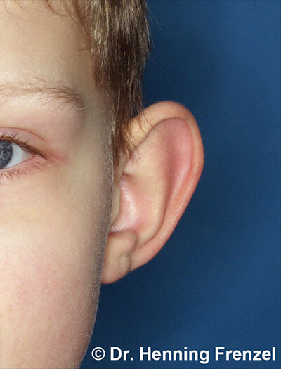
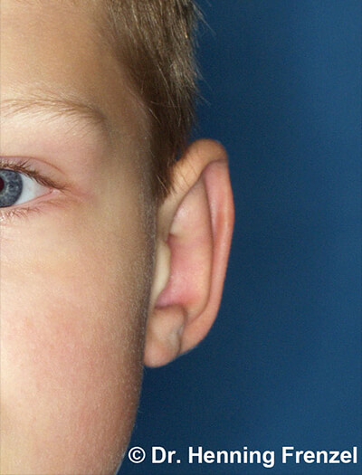
Ohrrekonstruktion – bei geringer Fehlbildung 1 Ohrrekonstruktion – bei geringer Fehlbildung 2 If the external ear is basically present and there is only a tiny defect, or the shape is malformed, it can be reconstructed through reshaping alone. The best known example of this is the correction of protruding ears. If the ears have a defect like, for example, tiny cup ears, this can be treated with a small local skin and cartilage graft.
Ear Reconstruction using a Prosthesis
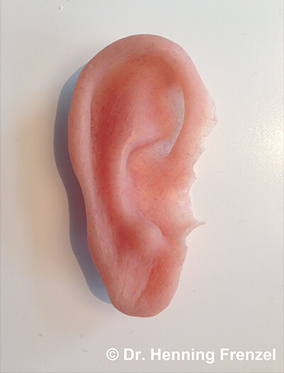
An artificial external ear is called an ear prosthesis. The missing ear is formed completely of silicon and is attached to the skull by magnets. The magnets are fixed in the skull under short anaesthesia.
The ear prosthesis itself is made by an independent maxillofacial prosthetist with whom we have a close working relationship.
The advantages of this method are that it is a less demanding procedure and that the appearance of the silicon ear resembles a normal ear. However, an artificial ear remains a foreign body. In addition the magnetic anchors penetrate the skin. We mainly use this method to treat acquired ear defects caused by tumours, injuries or unsuccessful ear reconstructions.
Future Outlook
There is intensive research into ear reconstruction using harvested cartilage. There are already promising signs. However they are not at the stage of general clinical use for patients.
